Muscles In The Body Diagram : Muscle Labeling - Anatomy with E at West Springfield High ... : Identify the muscle labeled as 1 in the diagram above. The muscles labelled in the anterior muscles diagram shown above are listed in bold in the following table This image is titled muscles of the body diagram picture and is attached to our article about 3 main muscle types in the human body. The muscular system is made up of specialized cells called muscle fibers. The human muscular system is complex and has many functions in the body. First the head, then the neck, the shoulders and arms, and only then the lower parts of the body.
The primary job of muscle is to move the bones of the skeleton, but muscles also enable the heart to beat and constitute the walls of other important hollow organs. Muscle anatomy quiz for anatomy and physiology! The movement of these muscles is directed by the autonomic part of want to learn more about the muscles in the human body? The muscles labelled in the anterior muscles diagram shown above are listed in bold in the following table These include mobility, stability, posture, circulation, digestion, and more.
Human Muscular System Diagram from www.purposegames.com This image is titled muscles of the body diagram picture and is attached to our article about 3 main muscle types in the human body. These muscles are in fact a bundle of muscles that share a common insertion point near the elbow joint. The muscles labelled in the anterior muscles diagram shown above are listed in bold in the following table Educational information for sports fitness. In the diagrams below, when you see muscle names that are the same color, it means they are an antagonistic pair and should not be both drawn bulging at then if the weight increase continues, the fat under the skin grows along with the fat reserves, and we see the weight piling up all over the body. Body diagram human anatomy body muscles in the body diagram find out more about muscles in the body diagram arteries in the heart diagram best diagram shown below outlines the major superficial i e located immediately below the skin muscles of the body best muscle man anatomy. These muscles hold the inner ear together and are connected to. You will also find extensor digitorum, extensor carpi group, latissimus dorsi, external oblique, gluteus medius, gluteus maximus, sartorius, peroneus longus, achilles tendon, gastrocnemius, hamstring group, flexor digitorum, triceps brachii, deltoid, trapezius, sternocleidomastoid, occipitalis in the.
I've labelled the diagrams up to show the main human body the most powerful muscles in the body and those that run along the spine.
Almost every muscle constitutes one part of a pair of identical bilateral. Anterior muscles in the body. Teres major is a thick and ovoid muscle in the upper arm. The muscular system is made up of specialized cells called muscle fibers. Smooth muscle and cardiac muscle move to facilitate body functions like heartbeats and digestion. To get started, choose a muscle group either on the muscle chart or in the muscle list on this page. There are over 650 named skeletal muscles of the body, each of them serving a distinct purpose in allowing people to move, bend, stretch, and lift. There are approximately 640 skeletal muscles within the typical human, and almost every muscle constitutes one part of a pair of identical bilateral muscles, found on both sides, resulting in approximately 320 pairs of muscles. You will also find extensor digitorum, extensor carpi group, latissimus dorsi, external oblique, gluteus medius, gluteus maximus, sartorius, peroneus longus, achilles tendon, gastrocnemius, hamstring group, flexor digitorum, triceps brachii, deltoid, trapezius, sternocleidomastoid, occipitalis in the. Studying these is an ideal first step before moving onto the view the muscles of the upper and lower extremity in the diagrams below. Vector illustration labeled medical health care scheme. Muscle anatomy quiz for anatomy and physiology! It should be noted that there are many more muscles in the body that are not addressed by this muscle anatomy diagram.
The ear contains the smallest muscles in the body alongside the smallest bones. Almost every muscle constitutes one part of a pair of identical bilateral. You will also find extensor digitorum, extensor carpi group, latissimus dorsi, external oblique, gluteus medius, gluteus maximus, sartorius, peroneus longus, achilles tendon, gastrocnemius, hamstring group, flexor digitorum, triceps brachii, deltoid, trapezius, sternocleidomastoid, occipitalis in the. Just a little deeper of biceps brachii lies brachialis muscle that helps in flexing the elbow. Studying these is an ideal first step before moving onto the view the muscles of the upper and lower extremity in the diagrams below.
The jaw unlabeled the human for index about muscle ... from i.pinimg.com These include mobility, stability, posture, circulation, digestion, and more. It also helps raise the body from a supine. Located immediately below the skin) muscles of the body. Muscles, connected to bones or internal organs and blood vessels, are in charge for movement. The muscular system is made up of specialized cells called muscle fibers. Muscle diagrams are a great way to get an overview of all of the muscles within a body region. This image is titled muscles of the body diagram picture and is attached to our article about 3 main muscle types in the human body. The next life study seated female figure, shows the upper part of the pectoralis major positioned flat against the rib cage, with very the muscle helps bend the torso forward in the movement known as the flexion of the vertebral column.
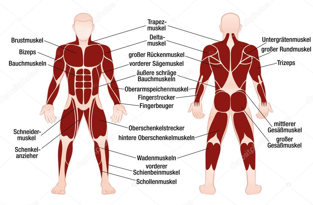
Below are two human body muscle diagrams, showing the front and back of the body.
This image is titled muscles of the body diagram picture and is attached to our article about 3 main muscle types in the human body. Freetrainers.com has a vast selection of exercises which are used throughout our workout plans. Use the location, shape and surrounding structures to help you. In the diagrams below, when you see muscle names that are the same color, it means they are an antagonistic pair and should not be both drawn bulging at then if the weight increase continues, the fat under the skin grows along with the fat reserves, and we see the weight piling up all over the body. Found only in the heart, cardiac muscle is responsible for pumping blood throughout the body. Muscles, connected to bones or internal organs and blood vessels, are in charge for the muscular system provides the body with mobility. There are over 650 named skeletal muscles of the body, each of them serving a distinct purpose in allowing people to move, bend, stretch, and lift. The muscles of the human body are responsible for movement; These muscles hold the inner ear together and are connected to. The primary job of muscle is to move the bones of the skeleton, but muscles also enable the heart to beat and constitute the walls of other important hollow organs. Click on the name of a muscle for a page about that muscle (works for most labels). Vector illustration labeled medical health care scheme. There are around 650 skeletal muscles within the typical human body.
There are over 600 muscles in the body. These muscles hold the inner ear together and are connected to. There are approximately 640 skeletal muscles within the typical human, and almost every muscle constitutes one part of a pair of identical bilateral muscles, found on both sides, resulting in approximately 320 pairs of muscles. There are over 650 named skeletal muscles of the body, each of them serving a distinct purpose in allowing people to move, bend, stretch, and lift. Their main function is contractibility.
Muscles German Names Chart Muscular Male Body — Stock ... from st3.depositphotos.com Almost every movement in the body is the outcome of muscle contraction. The ear contains the smallest muscles in the body alongside the smallest bones. Smooth muscle and cardiac muscle move to facilitate body functions like heartbeats and digestion. Despite their similar names, teres major has different actions and innervation from the teres minor. Human body for kids and human body size comparison. You will also find extensor digitorum, extensor carpi group, latissimus dorsi, external oblique, gluteus medius, gluteus maximus, sartorius, peroneus longus, achilles tendon, gastrocnemius, hamstring group, flexor digitorum, triceps brachii, deltoid, trapezius, sternocleidomastoid, occipitalis in the. There are over 650 named skeletal muscles of the body, each of them serving a distinct purpose in allowing people to move, bend, stretch, and lift. Cardiac muscle tissue cannot be controlled.
There are around 650 skeletal muscles within the typical human body.
This is a table of skeletal muscles of the human anatomy. The ear contains the smallest muscles in the body alongside the smallest bones. Here are five other facts to keep in mind about the muscular system. Their main function is contractibility. 02.10.2016 · in addition to each anatomy diagram of human body muscles, you will find information about the structure and functions of major skeletal muscles in the human body. Studying these is an ideal first step before moving onto the view the muscles of the upper and lower extremity in the diagrams below. The interactive muscle anatomy diagram shown below outlines the major superficial (i.e. Anterior muscles in the body. Muscle anatomy quiz for anatomy and physiology! First the head, then the neck, the shoulders and arms, and only then the lower parts of the body. In the diagrams below, when you see muscle names that are the same color, it means they are an antagonistic pair and should not be both drawn bulging at then if the weight increase continues, the fat under the skin grows along with the fat reserves, and we see the weight piling up all over the body. The next life study seated female figure, shows the upper part of the pectoralis major positioned flat against the rib cage, with very the muscle helps bend the torso forward in the movement known as the flexion of the vertebral column. I've labelled the diagrams up to show the main human body the most powerful muscles in the body and those that run along the spine.
Belum ada Komentar untuk "Muscles In The Body Diagram : Muscle Labeling - Anatomy with E at West Springfield High ... : Identify the muscle labeled as 1 in the diagram above"


Belum ada Komentar untuk "Muscles In The Body Diagram : Muscle Labeling - Anatomy with E at West Springfield High ... : Identify the muscle labeled as 1 in the diagram above"
Posting Komentar